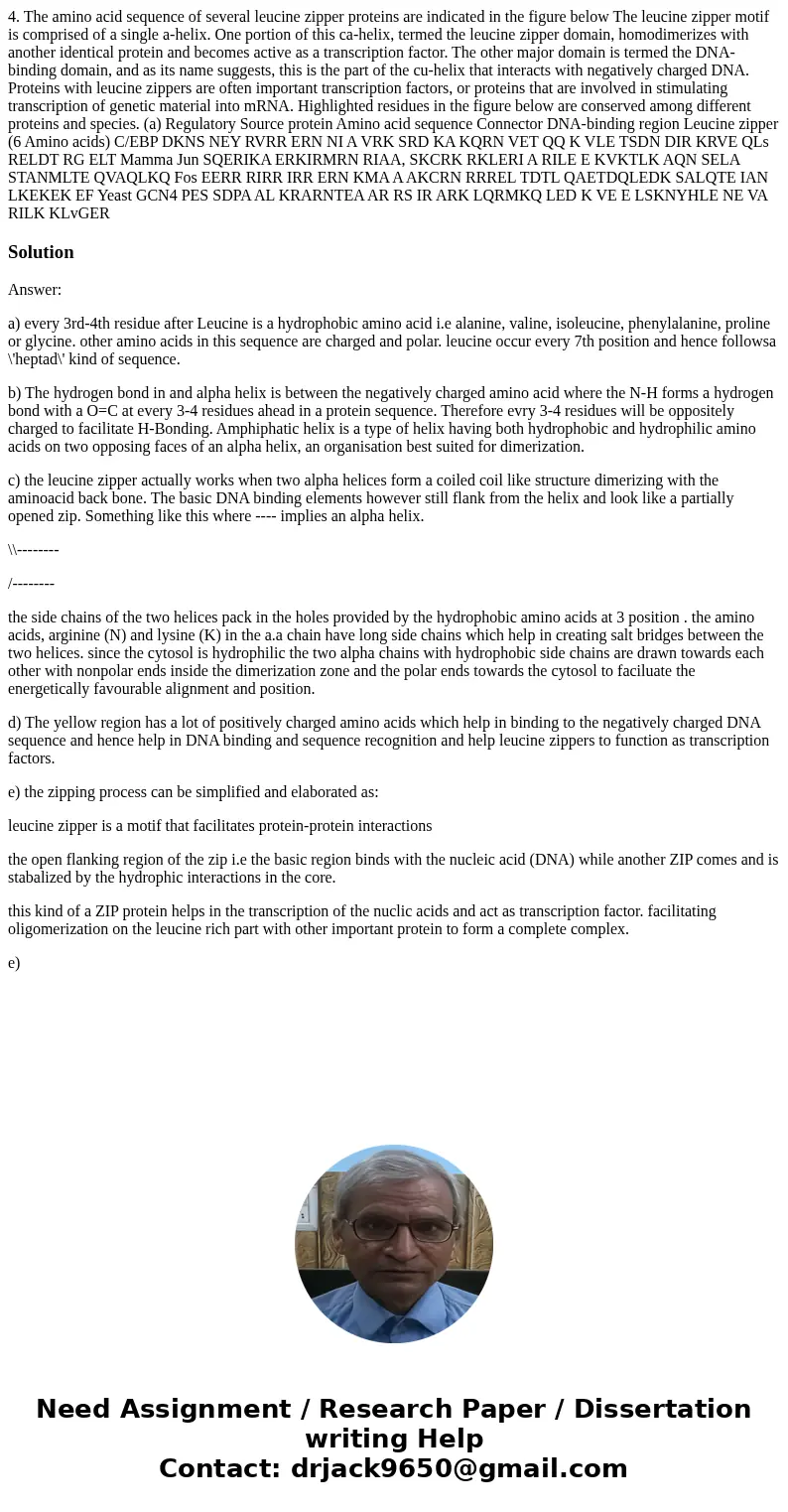
4 The amino acid sequence of several leucine zipper proteins
Solution
Answer:
a) every 3rd-4th residue after Leucine is a hydrophobic amino acid i.e alanine, valine, isoleucine, phenylalanine, proline or glycine. other amino acids in this sequence are charged and polar. leucine occur every 7th position and hence followsa \'heptad\' kind of sequence.
b) The hydrogen bond in and alpha helix is between the negatively charged amino acid where the N-H forms a hydrogen bond with a O=C at every 3-4 residues ahead in a protein sequence. Therefore evry 3-4 residues will be oppositely charged to facilitate H-Bonding. Amphiphatic helix is a type of helix having both hydrophobic and hydrophilic amino acids on two opposing faces of an alpha helix, an organisation best suited for dimerization.
c) the leucine zipper actually works when two alpha helices form a coiled coil like structure dimerizing with the aminoacid back bone. The basic DNA binding elements however still flank from the helix and look like a partially opened zip. Something like this where ---- implies an alpha helix.
\\--------
/--------
the side chains of the two helices pack in the holes provided by the hydrophobic amino acids at 3 position . the amino acids, arginine (N) and lysine (K) in the a.a chain have long side chains which help in creating salt bridges between the two helices. since the cytosol is hydrophilic the two alpha chains with hydrophobic side chains are drawn towards each other with nonpolar ends inside the dimerization zone and the polar ends towards the cytosol to faciluate the energetically favourable alignment and position.
d) The yellow region has a lot of positively charged amino acids which help in binding to the negatively charged DNA sequence and hence help in DNA binding and sequence recognition and help leucine zippers to function as transcription factors.
e) the zipping process can be simplified and elaborated as:
leucine zipper is a motif that facilitates protein-protein interactions
the open flanking region of the zip i.e the basic region binds with the nucleic acid (DNA) while another ZIP comes and is stabalized by the hydrophic interactions in the core.
this kind of a ZIP protein helps in the transcription of the nuclic acids and act as transcription factor. facilitating oligomerization on the leucine rich part with other important protein to form a complete complex.
e)

 Homework Sourse